Search This Blog

Sunday, March 3, 2013

Oral and Sublingual Immunotherapy as Treatment for Food Allergy and Anaphylaxis


An allergic response in the human body begins upon first exposure to the allergen.  (National Institute of Allergy and Infectious Diseases, 2013)  Upon the advent of the allergen, the immune system creates one type of antibodies, known as specific immunoglobulin E, with specific affinity to that substance.  (Lerner, 2010; National Institute of Allergy and Infectious Diseases, 2013.  For a diagram of an antibody, see image 1.)  A group of immunological molecules known as interleukins promote the cloning of these IgE antibodies.  (Robinson, 2013)  During the second exposure, there is a much larger immune response and elevated production flood the bloodstream with allergen-specific IgE molecules.  (Robinson, 2013)  These antibodies move throughout the bloodstream and attach to the antigen binding cites of specialized immune cells known as mast cells and basophils. (Urry, 2011; National Institute of Allergy and Infectious Diseases, 2013; Urry, 2011) IgE levels are typically very low in the bloodstream non-allergic person, but during an allergic reaction, the body begins producing excessive amounts of the allergen specific antibody.  (de Weck, 2012; Cohen, 2012)  The IgE molecules do not remain in the bloodstream for a long period of time.  (Cohen, 2012) Instead, immediately after its proliferation, the substance binds very strongly to the membrane of mast cells in bodily tissue and blood basophils, both of which are unique types of immune system inflammatory cells.  (Tsai, 2012; de Weck, 2012; National Institute of Allergy and Infectious Diseases, 2013)  As inflammatory cells, both mast cells and blood basophils contain “inflammatory mediators,” most commonly histamine and serotonin.  (de Weck, 2012)  The strong binding of IgE molecules to the high-affinity receptors of the inflammatory cells causes cross-linking between adjacent IgE molecules.  (Tsai, 2012; Cohen, 2012)  This cross-linking triggers a series of biochemical reactions and cascades within the mast cells that eventually result in the cell’s “degranulation.” (Tsai, 2012)  When the cell becomes “degranulated” its membrane bursts and massive amount of granule-associated mediators are released.  (Parker, 2007; Tsai, 2012)  These granule-associated mediators, or inflammatory mediators, are liable for the majority of signs and symptoms associated with allergic reactions.  (Tsai, 2012)  The most common granule-associated mediator in an allergic response is the molecule histamine, which induces inflammation in various tissues, although a variety of other molecules can be produced throughout the course of the reaction. (de Weck, 2012; Cohen, 2012)  Histamine induces dilation and increased permeability of small blood vessels in various body tissues and constriction of the bronchi.  (Robinson, 2013)  These symptoms result in fluid loss and swelling of the tissues.  (Robinson, 2013)  For diagrams of the cell response to IgE, see images 2-4.

Though the cause of food allergy is unknown, the disorder’s effects on the body are well documented and well understood.  (Staff, Mayo Clinic, 2011)  The mast cells, primary proliferators of an allergic reaction, are most common in the gastrointestinal tract, respiratory tract, and skin.  (Cohen, 2012)  Therefore, it is unsurprising that these areas are the most common sites of allergic reaction in the human body.  (Cohen, 2012)  Though allergic symptoms vary widely between individuals, a handful of them are common to most people who have food allergies.  These include, itching of the mouth, swelling of the lips and the tongue, symptoms that affect the gastrointestinal tract, including vomiting, diarrhea, abdominal cramps, and abdominal pain, hives, eczema and other skin issues, constricted throat or breathing, and a drop in blood pressure.  (National Institute of Allergy and Infectious Diseases, 2013)  The inflammatory mediators released by the affected cells bring on these archetypal symptoms.  (Tsai, 2012)  If the reaction is very severe, it can trigger a response known as anaphylaxis.  This response is marked by an incredibly wide range of symptoms, most notably, though, are swelling throughout the body tissues, wheezing, weak pulse, shock, and fainting.  (National Institute of Allergy and Infectious Diseases, 2013) Such reactions are frightening, unexpected, and have the potential to be deadly.  Children and adults who live with this condition have constant anxiety about anaphylaxis and other severe reactions from ingestion and contamination.  (Fleischer, 2013)  Allergies are the single most cause of days missed from school and work, and studies have shown that food allergy has significant effects on the social activities, meal preparation, and psychological state of children impacted by the condition.  (Flesicher, 2013; Bollinger, 2010; Lerner, 2010)  Though some children eventually grow out of their allergies as they age, many do not, especially those with peanut allergy.  (Cohen, 2012; Fleicher, 2013)

Currently, there are no cures for food related allergies.  (Fleischer, 2013)  For many other types of allergy, subcutaneous shots containing progressively higher amounts of the allergen have been shown to desensitize the patient and often reduce allergy symptoms.  (Fleischer, 2013)  However, when trials of subcutaneous injections were conducted on patients with peanut allergy, many individuals had adverse reactions, and the practice was deemed unsafe.  (Fleischer, 2013)  At the moment, patients with food allergies are advised to practice strict avoidance of the dangerous food.  (Cohen, 2012)  This can be very difficult, and there is a high likelihood of possible cross-contamination of foods in cafeterias and other public dining facilities.  If an individual’s food allergies are very mild, his or her symptoms can be treated with drugs known as antihistamines.  (Staff, Mayo Clinic, 2011)  These drugs negate the more mild effects of histamine and reduce the symptoms of the condition.  More severe allergic reactions and anaphylactic reactions are treated with epinephrine injections and trips to the emergency room.  (Staff, Mayo Clinic, 2011)  Epinephrine counteracts the symptoms of high levels of histamine, increasing blood vessel diameter, reduces blood vessel permeability, and relaxes the bronchi.  (Robinson, 2013)  Yet, these treatments are only temporary.  They treat the symptoms of allergies but they do not remedy the underlying intolerance that causes the reaction. 

For years, the means by which to remedy allergic intolerance has eluded scientific researchers.  There have been no broadly available therapeutic options for allergy suffers, and there have been myriad severe and fatal anaphylactic reactions brought on by contact with food.  (Fleischer, 2013) At long last, however, there is hope for those who suffer from these allergies, which comes in the form of oral and sublingual immunotherapy.  Oral and sublingual immunotherapies, first studied more than 100 years ago, have only recently come to the forefront of scientific research.  (Nowak-Wegrzyn, 2011)  During oral and sublingual immunotherapy, patients are administered small doses of the allergic food, either mixed into other non-allergic foods or under the tongue in extract form, respectively.  (Nowak-Wegrzyn, 2011)  The amount of allergens in these doses is gradually increased over the course of many weeks, resulting in an elevated tolerance to the substance in question. (Nowak-Wegrzyn, 2011) Patients beginning treatment first establish the amount of allergen they can consume without inducing a reaction.  (Fleischer, 2013)  After the initially dosage is established, each individual begins a series of daily build-up doses, each marginally greater than the last.  (Burks, 2012)  Patients periodically test their resistance to the allergen during “food challenges” wherein an individual consumes incremental amounts of the substance in order to determine his or her tolerance. (Fleischer, 2013)  The dosages can continue as long as the individual desires, or until an adverse reaction occurs.  (Burks, 2012)  The immunotherapy trains the body, through repeated expose, to tolerate what it had once rejected.  (Fessenden, 2012)
Though oral and sublingual immunotherapy procedures are still in trial phases, the results have been promising.  (Fleischer, 2013)  Sublingual immunotherapy treatment has been shown to raise tolerance in patients allergic to kiwi, hazelnut, peach, milk, and, most recently, peanut.  (Fleischer, 2013)  In individuals allergic to peanut, the most deadly known food allergy, sublingual immunotherapy raised tolerance from less than two grams of peanut protein to over ten grams in some cases.  (Fleischer, 2013)  Tolerance increased greatly when therapy was continued over many more weeks.  (Fleischer, 2013)  A similar study of oral immunotherapy conducted with egg protein was even more successful, with 70% of participants able to consume a cumulative dose of five grams of powder at ten months of treatment.  (Burks, 2012)  Oral and sublingual immunotherapy show great promise as food allergy treatment.  Measurements of the immune components of the subjects also revealed encouraging results.  In studies of the peanut specific antibody levels in immunotherapy patients, trial participants were found to have a decreased range of allergen-specific IgE molecules and greater levels of polyclonal allergen-specific IgG4 serum.  (Vickery, 2012; Burks, 2013; Fessenden, 2012)  IgG4 is another antibody, the serum of which is essential to the promotion of IgE in allergic reactions.  (Vickery, 2012)  There was also a decrease in peanut-specific interleukin production; interleukins, which promote the propagation of IgE molecules, are key to a strong allergic response.  (Blumchen, 2010; Robinson, 2013)  All of these biochemical signs correlate with a reduction or possible discontinuation of allergic response.  Furthermore, the treatment was found to be incredibly safe.  (Hofmann, 2009)  Very few individuals had adverse reactions, and those reactions were always in the hospital during build up days, where they could be safely treated.  (Hofmann, 2009) Unlike subcutaneous immunotherapy, which was discontinued because it was too dangerous for many patients, oral and sublingual immunotherapy treatments seem to be safe, even for those with serious peanut allergies.  (Blumchen, 2010; Hoffman, 2009)

Food allergy is a serious medical condition that affects approximately 20% of Americans today.  (Lerner, 2010)  For many years, individuals with the condition have lived in fear of anaphylactic shock and severe reaction.  (National Institute of Allergy and Infectious Diseases, 2013) A cure, other than symptomatic treatment, has long evaded researches and medical practitioners.  (Burks, 2012)  Recently, however, oral and sublingual immunotherapy have risen to prominence.  (Nowak-Wegryzn, 2011)  By slowly increasing consumption of allergen, these techniques increase body tolerance to the offending substance.  (Fleischer, 2013)  The treatment results in decreased diversity of allergen specific IgE and increased levels of allergen specific IgG4, both of which are good indicators of decreased allergic response.  (Vickery, 2012)  The treatment was found to be both safe and very effective.  (Hoffman, 2013; Fleischer, 2013)  It seems that the future advancements for allergy suffers lie in the very substances to which they are allergic.  As oral and sublingual immunotherapy gain prevalence and become widely used, one day we may see a world without food allergy. 

Appendix
Image 1
Image from the Biology 570 Immunology PowerPoint


Image 2
This image gives a basic overview of the IgE propagation, binding, and subsequent histamine release in allergic tissue

Lewis, Ricki. An Allergic Reaction - Overview. McGraw-Hill Companies, Inc, Digital Image. Nutri-Living, nd. Web. 25 Feb. 2013. <http://dft.ba/-allergicreaction>.


Image 3
This image shows the specific IgE molecules on an immune effector cell, and their cross-linking in the presence of an allergen.

Nature Reviews. Allergen Activation and Cross-Linking of IgE molecules. Digital image. Nature. Nature Reviews, n.d. Web. 25 Feb. 2013. <http://www.nature.com/nrd/journal/v3/n7_supp/images/nrd1408-f1.jpg>.


Image 4
An image displaying the various paths of mast-cell activation.  Note the presence of IgE and allergens, as well as the granule-associated mediators leading to inflammation of the tissue.


Mast Cell Activation. Digital image. AccessScience. McGraw-Hill Education, 2012. Web. 25 Feb. 2013. <http://www.accessscience.com/content/Mast%20cells/900114>.

Bibliography
Blumchen, Kathrina, Md, Helen Ulbricht, Ute Staden, MD, Kerstin Dobberstein, John Beschorner, Lucila C. L. De Oliveira, MD, Wayne G. Shreffler, MD, PhD, Hugh A. Sampson, MD, Bodo Niggemann, MD, Ulrich Wahn, MD, and Kirsten Beyer, MD. "Oral Peanut Immunotherapy in Children with Peanut Anaphylaxis." Journal of Allergy and Clinical Immunology 126.1 (2010): 83-91. ScienceDirect. Mosby, Inc., July 2010. Web. 25 Feb. 2013. <http://www.sciencedirect.com/science/article/pii/S0091674910007268>.

Bollinger, Mary E., DO, Lynnda M. Dahlquist, PhD, Kim Mudd, RN, MSN, Claire Sonntag, BA, Lindsay Dillinger, BA, and Kristine McKenna, MS. "The Impact of Food Allergy on the Daily Activities of Children and Their Families." Annals of Allergy, Astma, & Immunology 96.3 (2006): 415-21.ScienceDirect. Elsevier Inc., 18 Feb. 2010. Web. 26 Feb. 2013. <http://www.sciencedirect.com/science/article/pii/S1081120610609088>.

Burks, A. W., MD, Stacie M. Jones, MD, Robert A. Wood, MD, David M. Fleischer, MD, Scott H. Sicherer, MD, Robert W. Lindblad, MD, Donald Stablein, PhD, Alice K. Henning, MS, Brian P. Vickery, MD, Andrew H. Liu, MD, Amy M. Scurlock, MD, Wayne G. Shreffler, MD, PhD, Marshall Plaut, MD, and Hugh A. Sampson for the Consortium of Food Allergy Research (CoFAR), MD. "Oral Immunotherapy for Treatment of Egg Allergy in Children."The New England Journal of Medicine (2012): 233-43. The New England Journal of Medicine. Massachusetts Medical Society, 19 July 2012. Web. 25 Feb. 2013. <http://www.nejm.org/doi/full/10.1056/NEJMoa1200435>.

Cohen, John J. "Food Allergy." AccessScience. McGraw-Hill Education, 2012. Web. 22 Feb. 2013. <http://www.accessscience.com/content.aspx?searchStr=Food+Allergy&id=802770>.
De Weck, A. L. "Allergy." AccessScience. McGraw-Hill Eductionation, 2012. Web. 23 Feb. 2013. <http://www.accessscience.com/content/Allergy/024150>.

Fessenden, Marissa. "The Exposure Cure." Scientific American 16 (2012): 307. Nature. Scientific American, 18 Sept. 2012. Web. 25 Feb. 2013. 
<http://www.nature.com/scientificamerican/journal/v307/n4/full/scientificamerican1012-16.html>.

Fleischer, David M., MD, A. W. Burks, MD, Brian P. Vickery, MD, Amy M. Scurlock, MD, Robert A. Wood, MD, Stacie M. Jones, MD, Scott H. Sicherer, MD, Andrew H. Liu, MD, Donald Stablein, PhD, Alice K. Henning, MS, Lloyd Mayer, MD, Robert Lindblad, MD, Marshall Plaut, MD, Hugh A. Sampson, MD, and Consortium of Food ALlergy Research (CoFAR). "Sublingual Immunotherapy for Peanut Allergy: A Randomized, Double-Blind, Placebo-Controlled Multicenter Trial." Journal of Allergy and Clinical Immunology 131.1 (2013): 119-27. ScienceDirect. Elsevier Properties S.A., Jan. 2013. Web. 24 Feb. 2013. <http://www.sciencedirect.com/science/article/pii/S0091674912018246>.

Hine, Robert. "Allergy." Science Online. Facts On File, Inc., n.d. Web. 25 Feb. 2013. <http://www.fofweb.com/activelink2.asp?ItemID=WE40&SID=5&iPin=FDBF0094&SingleRecord=True>.

Hofman, Alison M., MD, Amy M. Scurlock, MD, Stacie M. Jones, MD, Kricia P. Palmer, MD, Yuliua Lokhnygina, PhD, Pamela H. Stelle, CPNP, Jamet Kamilaris, RN, and A. W. Burks, MD. "Safety of a Peanut Oral Immunotherapy Protocol in Children with Peanut Allergy." Journal of Allergy and Clinical Immunology 124.2 (2009): 286-91. ScienceDirect. Elsevier B.V., 27 May 2009. Web. 27 Feb. 2013. <http://www.sciencedirect.com/science/article/pii/S0091674909005569>.

Lerner, K. L., and Brenda W. Lerner. "Allergy." The Gale Encyclopedia of Science. Gale Cengage Learning, 2010. Web. 25 Feb. 2013. <http://ic.galegroup.com/ic/scic/ReferenceDetailsPage/ReferenceDetailsWindow?failOverType=&query=&prodId=SCIC&windowstate=normal&contentModules=&mode=view&displayGroupName=Reference&limiter=&currPage=&disableHighlighting=false&displayGroups=&sortBy=&source=&search_within_results=&action=e&catId=&activityType=&scanId=&documentId=GALE%7CCV2644030067>.

Mast Cell Activation. Digital image. AccessScience. McGraw-Hill Education, 2012. Web. 25 Feb. 2013. <http://www.accessscience.com/content/Mast%20cells/900114>.

National Institute of Allergy and Infectious Diseases. "Food Allergy: An Overview." U.S. Department of Health and Human Services, 2013. Web. 11 Jan. 2013. <http://www.niaid.nih.gov/topics/foodallergy/documents/foodallergy.pdf>.

Nature Reviews. Allergen Activation and Cross-Linking of IgE molecules. Digital image. Nature. Nature Reviews, n.d. Web. 25 Feb. 2013. <http://www.nature.com/nrd/journal/v3/n7_supp/images/nrd1408-f1.jpg>.

Nowak-Wegrzyn, Anna, MD, and Hugh A. Sampsom, MD. "Future Therapies for Food Allergies." Journal of Allergy and Clinical Immunology 127.3 (2011): 558-73. ScienceDirect. Mosby, Inc., Mar. 2011. Web. 26 Feb. 2013. <http://www.sciencedirect.com/science/article/pii/S0091674911000030>.

Parker, Steve. "Allergies." The Human Body Book. 1st ed. New York: DK Pub., 2007. 166. Print.

Robinson, Keith A. "Biology 570 Immunology." Massachusetts, USA, Andover. 26 Feb. 2013. Lecture.

Staff, Mayo Clinic. "Food Allergy." Mayo Clinic. Mayo Foundation for Medical Education and Research, 11 Feb. 2011. Web. 27 Jan. 2013.

Tsai, Mindy. "Mast Cells." AccessScience. McGraw-Hill Education, 2012. Web. 23 Feb. 2013. <http://www.accessscience.com/content/Mast%20cells/900114>.

Urry, Lisa A., Michael L. Cain, Steven A. Wasserman, Peter V. Minorsky, and Robert B. Jackson. "Allergies." Campbell Biology AP Edition. By Jane B. Reece. 9th ed. San Francisco: Pearson Benjamin Cummings, 2011. 947. Print.

Varsheney, Pooja, MD, Stacie M. Jones, MD, Amy M. Scurlock, MD, Tamara T. Perry, MD, Alex Kemper, MD, MPH, MS, Pamela Steele, CPNP, Anne Hiegel, RN, Jamet Kamilaris, RN, Suzanne Carlisle, RN, Xiaohong Yue, MS, Mike Kulis, PhD, Laurent Pons, PhD, Brian Vickery, MD, and A. W. Burks, MD. "A Randomized Controlled Study of Peanut Oral Immunotherapy: Clinical Desensitization and Modulation of the Allergic Response."Journal of Allergy and Clinical Immunology 127.3 (2011): 654-60. ScienceDirect. Mosby, Inc., Mar. 2011. Web. 25 Feb. 2013. <http://www.sciencedirect.com/science/article/pii/S0091674911000509>.

Vickery, Brian P., MD, Jing Lin, PhD, Micheal Kulis, PhD, Zhiyan Fu, PhD, Pamela H. Steele, MSN, CPNP, Stacie M. Jones, MD, Amy M. Scurlock, MD, Gustavo Gimenez, BSc, Ludmilla Bardina, MSc, Hugh A. Sampson, MD, and A. W. Burks, MD. "Peanut Oral Immunotherapy Modifies IgE and IgG4 Responses to Major Peanut Allergens." Journal of Allergy and Clinical Immunology 3rd ser. 131.1 (2013): 128-34. ScienceDirect. Mosby Inc., 27 Nov. 2012. Web. 11 Jan. 2013. <http://www.sciencedirect.com/science/article/pii/S0091674912017721>.

Saturday, January 5, 2013

Tissue Types: Animal Structure and Function

The human body is a remarkably complex organization of substances, from cells to organs.  Essential to the makeup of the body are tissues.  There are four different main types of tissues in the body, each with several sub-types.

The first tissue type is the epithelial tissue, also commonly called the epithelia.  This tissue is made up of many layers of cells.  It makes up the skin, which covers the outside of the body, and covers organs and cavities within the body to protect them from damage.  This tissue type has five common sub-types:

1. The stratified squamous epithelium.

The stratified squamous epithelium, shown to the left, is a multi-layered tissue.  The most notable aspect of this tissue is the speed at which its cells are capable of replicating themselves.  New cells are formed at the base of the tissue and these cells rise to the top as old, dead cells are worn off.  This epithelial tissue is present on areas of the body that are frequently exposed to wear and the outside world, such as the skin.


2.  The pseudostratified columnar epithelium.

This tissue type is composed of one layer of cells that are a variety of heights, but whose nuclei look similar to those of the stratified epithelia.  This tissue type is most easily recognizable by the cilia on its surface, though pseudo stratified columnar epithelium can lack cilia in some cases.  This tissue often makes up a membrane of mucous (moved by the cilia) in the respiratory tract.


3.  The simple squamous epithelium.


The simple squamous epithelium is a single layer of large, flat cells.  It is the thinnest of any epithelial tissue.  This tissue type is notable for the incredibly fast rate at which substances can diffuse through it (owing to the fact that it is a single layer of very thin cells).  This tissue type is found in areas where diffusion of substances is essential, such as the lungs or the Bowman's capsule of the kidney.


4.  The simple columnar epithelium

The simple columnar epithelial tissue is made up of very large, long, rectangular cells, as shown in the image on the left.  These cells are key in absorption, particular in areas that are subject to abrasion and wear (i.e. the intestines and the digestive tract).  They also protect the rest of the body from any harmful bacteria that might have found there way into these areas by secreting a mucous coat, which is kept fluid by many microvilae.


5.  The cuboidal epithelium
The cuboidal epithelium is named for the cube-like cells that make up the tissue (shown on the left).  These cells are also key to secretion.  This tissue surrounds various structures within the body to protect from pathogens and wear and tear.  They are found in the kidneys and many glands within the body, where they assist with absorption.


The next tissue type is connective tissue.  Quite simply, this is the tissue that connects very many parts of the body.  This tissue makes up a matrix that holds the internal organs in place.  The matrix contains many cells, including fibroblasts and macrophages.  There are three different varieties of fiber that compose the connective tissue: collagenous fibers, reticular fibers, and elastic fibers.

1.  Loose connective tissue

The loose connective tissue, made up of a loose network of collagenous fibers and elastic fibers, is the most abundant connective tissue in the human body.  It binds a variety of structures together (i.e. connecting muscle fibers) and helps to make up layers of the skin.


2.  Fibrous connective tissue

The fibrous connective tissue, found most typically in tendons and ligaments, is a dense tissue made up primarily of collagenous fibers.  These fibers are made very small, strong collagen fibrils.  Interestingly enough, these collagen fibers are fluorescent under a UV light.


3.  Bone
Vertebrates have skeletons made up of bone tissue.  Bone is a very hard tissue, created by osteoblasts which initially produce a strong matrix of collagen.  Then calcium,magnesium, and phosphate ions mix together inside of the matrix, forming a hard mineral substance.  The ring shapes shown on the left are called "osteons" and are made up of circular layers of the matrix surrounding a passage of blood vessels and nerves.


4.  Adipose tissue
Adipose tissue is the substance more commonly known as "fat."  The cells make up a loosely connected tissue.  Each individual adipose cell stores fat droplets.  When fat is being stored the cells expand to hold more and when it is being used up they contract as the fuel is burned.  The adipose cells are found throughout out the body's matrix of connective tissue.  They are essential to well being.  They provide padding, insulate the human being to assist in maintaining body temperature, and help by storing energy in the form of fat that can be burned in times of famine or after great energy expenditure.


5.  Cartilage


Cartilage is a network of collagen-containing fibers within a mixture of proteins and carbohydrates commonly referred to as chondroitin sulfate.  Chondrocytes are the cells which create cartilage by producing both the collagen and the chondroitin sulfate.  The combination of these two substances is durable and pliable.  In fact, cartilage makes up the skeletons of many animals with vertebrae while they are still within the womb.  In an adult human it can be found throughout the body providing cushioning between bones, such as the knees and the vertebrae.


6.  Blood
Blood is the substance which flows throughout the vein sin the human body.  It is made up of plasma (a liquid ECM), erythrocytes (commonly known as red blood cells), leukocytes (known as white blood cells) and platelets.  Each of these substances serves a unique purpose.  The erythrocytes act as oxygen carriers, shuttling the substance throughout the body to be used in  cellular respiration.  The leukocytes are part of the immune system and defend the body from harmful pathogens.  The platelets allow the blood to clot, which is essential to healing cuts and abrasions.

The third tissue type is muscle tissue.  Muscle tissue is the tissue that allows animals and people to move.  It is composed if actin and myosin protein filaments that enable muscle contraction.

1.  Skeletal Muscle
Skeletal muscle, so named because it makes up the muscles that are attached to the skeleton, enables voluntary movement.  This muscle type is also known as striated muscle.  It is composed of muscle fibers (collections of long cells shown on the left).


2.  Smooth muscle


Smooth muscle differs from skeletal muscle in that it has no striations (this cal be seen by comparing the two images).  It is present in the digestive tract and other internal organs.  They are long and narrow, and cause involuntary movement, for example movement of the stomach and the arteries.


3.  Cardiac muscle

Cardiac muscle is the tissue that makes up large portions of the heart.  It is responsible for the heart beating.  The cells are interconnected and thereby organize their heart's "beats" or contractions within the contractile wall.


The fourth and final type of tissue is nervous tissue.  This is the tissue that makes up the nervous system.

1.  Neurons
Neurons are the most basic aspects of the nervous system.  It takes electrical impulses from the rest of the body's cells and the dendrites which stretch down from the neuron's body.  The transmit these impulses to each other or different cells through axons, which are gathered together to compose nerves.


2. Glial cells
The glial cells bring nutrients, provide insulation for, and restore the different neurons in the brain.  Occasionally they also monitor neuron function.


Sources:
http://en.wikipedia.org/wiki/Epithelium (accessed 1/5/13)
Campbell Biology Ninth Edition (Reece, Urry, Cain, Wasserman, Minorsky, Jackson) p. 856-859
http://en.wikipedia.org/wiki/Pseudostratified_columnar_epithelium (accessed 1/5/13)
http://www.bio.davidson.edu/people/kabernd/BerndCV/Lab/EpithelialInfoWeb/Simple%20Squamous%20Epithelium.html (accessed 1/5/13)
http://bio.rutgers.edu/~gb102/lab_6/601dm-simplecol.html (accessed 1/5/13)
http://www.bio.davidson.edu/people/kabernd/BerndCV/Lab/EpithelialInfoWeb/Simple%20Columnar%20Epithelium.html (accessed 1/5/13)
http://www.bio.davidson.edu/people/kabernd/BerndCV/Lab/EpithelialInfoWeb/Simple%20Cuboidal%20Epithelium.html (accessed 1/5/13)
http://bio.rutgers.edu/~gb102/lab_6/603bm-loose.html (accessed 1/5/13)
http://www.aps.uoguelph.ca/~swatland/ch2_3.htm (accessed 1/5/13)

Images courtesy of (in order as shown in the post):
http://www.nku.edu/~dempseyd/stratified%20squamous%20epithelium.gif
https://blogger.googleusercontent.com/img/b/R29vZ2xl/AVvXsEhoDxK_HV4c6Ncuj2Q9jteUPcZ6-_Ksc_73Je5We8znG15wSr7pyauGyN_ztnJ1H5g3w2dsmNaaweIjSpF-MigrQbzDHs9Cnchp7h99aDskg6Gra-l3436qKG5BNekTmHznJGcp4_U_Bis/s1600/Pseudostratified+columnar+epithelium+06.jpg
http://dft.ba/-simplesquamous
http://www.carlalbert.edu/assets/images/Math%20and%20Science/simple_columnar_epithelium.jpg
http://science.tjc.edu/Course/BIOLOGY/1409/cuboidal2.6-9.jpg
http://stevegallik.org/sites/histologyolm.stevegallik.org/images/areolar_01.jpg
http://washington.uwc.edu/about/wayne.schaefer/TISSUES/fibrous_connective_tissue_tendon.jpg
http://www.umm.edu/graphics/images/en/1679.jpg
http://3dsciencepics.com/wp-content/uploads/adipose_tissue.jpg
http://www.vetmed.vt.edu/education/curriculum/vm8054/labs/Lab7/IMAGES/Elastic%20cartilage%20photo%20EDITED.JPG
http://sciweb.hfcc.edu/Biology/AP/134/lab/lab%20guide%20images/Histology/Bio%20134%20photomicrographs.400x/Blood%20CT%20400x.1.jpg
http://stevegallik.org/sites/histologyolm.stevegallik.org/images/SkeletalMuscle_2.jpg
http://stevegallik.org/sites/histologyolm.stevegallik.org/images/Smooth%20muscle%2006.jpg
http://image.funscrape.com/images/c/cardiac_muscle-752.jpg
http://www.doctortipster.com/wp-content/uploads/2012/02/neuron-network.jpg
http://www.topnews.in/health/files/Glial-Cells-Neurons.jpg

Wednesday, November 28, 2012

The Science of Lying

Here's an excellent video by Hank Green of SciShow on the science of lying!

Monday, September 24, 2012

Everything You've Ever Wanted to Know About Carbohydrates!

Diagram of a glucose molecule.

You've probably heard quite a bit about carbohydrates.  When dieting you avoid them like the plague; they're in pastas, pizzas, and many other delicious Italian dishes.  But what, in fact, is a carbohydrate?  Carbohydrates are defined as molecules that are some form of sugar (either a polymer or monomer).  These sugars are composed of three elements:  Carbon, Hydrogen, and Oxygen in a 1:2:1 ratio. The carbohydrate most commonly found in nature is Glucose, with the molecular formula C6H12O6 (See right).  Carbohydrates perform several key functions for cells.  They store energy and they provide structure for living organisms.  The simplest sugars, such as Glucose, are referred to as monosaccharides.  Two monosaccharides can be combined to form a molecule called a disaccharide.  The process by which this occurs is called dehydration synthesis.  In dehydration synthesis, one monosaccharide donates a hydroxide ion (OH-) and another donates a hydrogen ion (H+).  These two ions form one molecule of H2O.
The result is that the two monosaccharides are held together in a glycosidic linkage.  Many monosaccharides can be combined in this way to form a polysaccharide.  Polysaccharides are the molecules that we usually think of as "carbohydrates".  They are composed of chains that have hundreds or even thousands of monosaccharides joined together via dehydration.  Interestingly enough, these chains can be broken apart using a process named hydrolysis.  Hydrolysis is, in essence, the opposite of dehydration.  In hydrolysis, the bond between two monosaccharides is broken by introducing a water molecule.  Energy is released, and the glycosidic linkage dissipates.  This is how carbohydrates store energy, by creating large polysaccharides when energy is abundant and breaking up the polysaccharides when energy is scarce.  

There are four common types of carbohydrates found in nature, described below:

1.  Starch

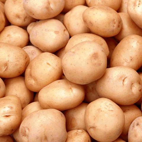
Potatoes, a common starch
Starch is an energy storage polysaccharide found in plants, specifically in their granules. (Want to know more about cell parts?  Click here.)  These molecules are spiral shaped, allowing for more efficient and compact storage.  The presence of starch enables a plant to stockpile its excess glucose and use it later for energy.  Starch can be broken apart by both humans and animals for energy.  Starch is, in fact, composed of two distinct substances.  The first is known as amylose.  Amylose, making up 20% of the molecule's composition, is soluble in water and has a linear shape.  Amylopectin makes up the other 80%.  Amylopectin is branched and, for the most part, not able to be dissolved by water.  Examples of starch include corn, rice, and potatoes.  

2. Glycogen

Glycogen is starch's animal counterpart.  Mostly found in the liver and muscle cells, glycogen is essential to an animal's well-being.  Without glycogen, normal body conditions cannot be maintained for long periods of time.  In fact, human beings must eat some kind of food with carbohydrates, otherwise glycogen stores will be depleted and muscle capabilities will be decreased.

3.  Cellulose

A cross-section of wood.

Cellulose provides structure to plants.  It is a straight, unbranched molecule.  Its components are held together by hydrogen bonds and mibrofibrils.  Cellulose is most commonly found in the cell walls of plants, and is the most commonly found organic compound on the planet.  Interestingly, cellulose is a polymer of glucose, with different glycosidic linkages than that of starch.  Hence, it has a flat shape while starch has a helix shape.  Cellulose is the main component in wood.  Some species of animals, such as termites, have special enzymes that enable them to digest this substance, however humans cannot digest it.


4. Chitin

Chitin is the structural carbohydrate found in many animals.  It is present in the exoskeletons of insects, and the cell walls of funguses.  Chiten has beta linkages with nitrogen attachments, creating its hard, tough surface.  


Sources:
Campbell Biology 9th AP Edition
http://www.medicalnewstoday.com/articles/161547.php
http://www2.chemistry.msu.edu/faculty/reusch/VirtTxtJml/carbhyd.htm
http://www.mansfield.ohio-state.edu/~sabedon/068dhsyn.gif
http://homebrewsake.com/wp-content/uploads/2010/04/glucose.gif
http://stemlynsblog.org/wp-content/uploads/2012/07/starch.jpg
http://upload.wikimedia.org/wikipedia/commons/thumb/0/0b/Taxus_wood.jpg/300px-Taxus_wood.jpg

Saturday, April 7, 2012

Taste - A Tongue in Cheek Sense

Picture courtesy of: dft.ba/-2luc
To function as human beings, we all need to eat. Food powers our bodies, allowing for the production of ATP (as previously mentioned on this site) which enables us to perform any and all of our activities, from breathing to dancing and thinking. Taste is essential to consumption. If food were tasteless, there would be little motivation to fuel our bodies. Therefore, we developed a way to enjoy various foods that would fuel our bodies.

On the top of one's tongue there is a collection of organs known as gustatory calyculi or, more commonly, taste buds. The tongue is home to approximately 100,000 individual taste buds. Each of these buds contains about 50-100 cells known as "taste cells" each of which, contrary to popular belief, can perceive a variety of flavors. These cells have a life span of 1-2 weeks before they die and are replaced. There are additional buds on the top, sides, and rear of the mouth, along with the throat. The gustatory calyculi are able to perceive five different types of flavor; bitter, sweet, salty, sour, and savory (often referred to as "umami," a Japanese word that means savory). 

Bitter
Some examples of bitter foods
The perception of bitter taste occurs on a certain receptor in the taste cell known as a G-protein-coupled receptor, specifically the taste receptors, type 2 (TAS2Rs). On these receptors, the substance combines with a substance known as gustducin. Then calcium ions (charged particles) depart from a section of the cell known as the endoplasmic reticulum. These calcium ions create a neurological response in the brain that triggers the "sensory neurons" in charge of perceiving the bitter taste. Interestingly enough, the tongue is more sensitive to bitter flavors than any others. Evolutionarily, this is most likely due to the fact that many toxic foods are quite bitter. Therefore, people more sensitive to this flavor would have been more likely to survive natural selection. Two man made substances, PTC (phenylthiocarbamide) and PROP (6-n-propylthiouracil) taste very bitter to people with specific genetic make ups, but are tastless to people with other genotypes. This is a common test to determine if one is a "supertaster" (more on this later).This ability to taste these chemicals is cause by just two alleles (at TAS2R38 location). 

Some examples of sweet foods, specifically candies
Sweet
Sweet food perception also occurs on G-protein-coupled receptors, specifically T1R2+3 and T1R3. It also involves combining the foodstuff with gustductin. How sweet the substance is perceived as depends on how tightly it binds with the two receptors. 


Salty
Table salt, or NaCl, commonly found in salty foods
The taste receptors for salt, specifically sodium chloride (NaCl) are channels through which sodium ions can flow into the cell. Calcium ions also enter the cell and again trigger a neurological response the causes the perception of the salty flavor. There is one hormone known as aldosterone that regulates the number of salt receptors in the mouth and also the levels of sodium in the body. Other substances that can trigger a similar response include KCl )potassium chloride), NH4+ (ammonium), and cations from the group of alkali earth metals.

Some sour fruits commonly found in nature
Sour
The flavor of sour foods is caused by their acidity. Acidity of a substance is relative to its number of H+ ions, so it is natural that these ions would be the cause of the sour perception. The taste cells perceive sour by expressing the protein PKD2L1. However, the H+ ions themselves can also illicit the taste response, so this gene is not required for this taste perception. 


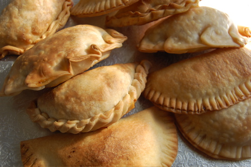
Some Savory Empanadas
Savory (Umami)
The perception of the umami taste comes from glutamic acid salts (for example, MSG, which is often found in chinese food). This once again occurs on the G-protein-coupled receptors, specifically the glutamate receptors, subsets T1R1 and T1R3. 

Supertasters
One of the most fascinating evolutionary phenomena associated with taste is that of the supertasters. It is believed that this anomaly is caused by an increased amount of taste buds present on the tongue. The first mention of the term "supertaster" was in a study conducted by Linda Bartoshuk. She was conducting a study on taste perception in the 1990s, when her research team noticed that some of the participants had increased response to tastes. As previously mentioned, the ability to taste the chemicals PTC and PROP have been linked with the ability to super taste. It appears that this specific taste receptor comes from one gene (TAS2R38). People who express this genotype prefer sweets as children, do not enjoy alcohol as much as their counterparts, less vegetable consumption, and a lessened tendency to smoke. It is estimated that 25% of people are are supertasters, 50% are regular tasters, and another 25% have a decreased number of taste buds. 

If you don't have access to PROP or PTC, here's a quick and easy test to see if you are a supertaster:

Step 1:
Using blue food color or a blue lollipop, stain your tongue blue. 

Step 2:
Place a piece of hole punched paper on your tongue, making sure that the hole is on top of the blue stained area.
The tongue of a supertaster

Step 3:
Look in the mirror. Using a magnifying glass, count the number of papillae you see. (Unlike the rest of your tongue, the papillae will be unstained). 

If there are more than 35, you are likely a supertaster. If there are 15-35, you are a medium taster, If there are less than 15, you are a non-taster.


Fun Facts:
-Women are more likely to be supertasters than men
-Asians, Africans, and South Americans are also more likely to be supertasters
-The bass player for the band Muckaferguson is a supertaster. They Might Be Giants wrote a song about him (John Lee Supertaster)
-Supertasters like salty foods
-Supertasters are less likely to enjoy fatty and sugary foods
-Supertasters are generally more skinny than regular tasters because they do not enjoy many unhealthy foods, however, they have elevated risk of colon cancer because they do not enjoy bitter vegetables

Still unsure if you're a supertaster or regular taster? Try this test, provided by the BBC. 

Sources:
http://users.rcn.com/jkimball.ma.ultranet/BiologyPages/T/Taste.html
http://faculty.washington.edu/chudler/tasty.html
http://en.wikipedia.org/wiki/Taste
http://en.wikipedia.org/wiki/Supertaster#Identifying_a_supertaster
http://www.todayifoundout.com/index.php/2010/06/how-to-tell-if-you-are-a-supertaster/
http://www.npr.org/templates/story/story.php?storyId=127914467
http://www.bbc.co.uk/science/humanbody/body/articles/senses/supertaster.shtml

Images:
http://hecooksshecooks.net/wp-content/uploads/2009/05/bitter-foods.jpg
http://www.virginmedia.com/images/ysp%20food%206%20sweets%20on%20la%20rambla,%20barcelona%20by%20helen%20sandell.jpg
http://whatscookingamerica.net/Information/Salt1.jpg
http://www.petitekitchenesse.com/wp-content/uploads/2010/10/fruit.jpg
http://joepastry.site.aplus.net/pics/empanadas1.jpg
http://blogsoop.com/blog/wp-content/uploads/2007/07/supertasters_supertaster.jpg

Sunday, February 5, 2012

That's Some Hungry Fungi! Fungus Capable of Eating Plastic Found.

Pestalotiopsis Microspore
Originally created in the 1900s, synthetic plastics have myriad uses. Plastic makes up disposable water bottles, bags, packaging, headphones, cellphones, and countless other everyday items. However, plastics are also detrimental to the environment. Incredibly durable and taking hundreds or even thousands of years to degrade, we waste alarmingly large amounts of the substance every year. The plastics that we throw out can sit in landfills for generations, seemingly impossible to get rid of. However, researchers at Yale have found a fungus from the Amazon rainforest with a rather fantastic property - it is capable of digesting polyurethane.

Known as Pestalotiopsis microspore (member of the family Amphisphaeriaceae, order Xylariales, class Sordariomycetes, subclass Xylariomycetidae, and phylum Ascomycota), the fugus was first found in Ecuador. It is able to subside solely on polyurethane and is the first fungus ever discovered that is capable of doing so. Not only is this remarkable in and of itself, but the fungus can also break down the plastic anaerobically (without oxygen). The team of scientists also found the enzyme that enables the fungus to digest the polyurethane. The researchers believe that this enzyme is a promising development in bioremediation and a huge leap forward in removing plastics from landfills.

Formation Reaction of Polyurethane 
Sources:
http://www.pcworld.com/article/249216/yale_discovers_a_fungus_that_eats_plastic.html
http://www.popsci.com/science/article/2012-02/rainforest-fungus-eats-plastic-potentially-solving-landfill-problems
http://en.wikipedia.org/wiki/Plastic#Environmental_issues
http://en.wikipedia.org/wiki/Pestalotiopsis_microspora

Images courtesy of:
http://www.jessicastuartmusic.com/wp-content/gallery/pre-jsfew/mushroom-spores.jpg
http://upload.wikimedia.org/wikipedia/commons/c/ca/Polyurethane.png


Wednesday, January 18, 2012

Study Reveals Ability of Yeast to Quickly Evolve

An article on today's New York Times chronicled a fascinating study at the University of Minnesota, a study that may change the way we study evolution. In the study, students discovered that yeast cells used to brew beer can form basic bodies in approximately two weeks.


Researchers created an experiment in which brewer's yeast was fed sugar and encouraged to reproduce, in hopes that the typically unicellular organism would perhaps create some kind of multicellular creature. To encourage this evolution the scientists raised so called "lines of yeast" from a single cell in 10 individual flasks full of "broth" - a soupy substance of nutrients. The flasks were shaken constantly for twenty four hours, and then allowed to settle. One drop of settled cells were put into a new flask and the yeast grew again. This meant that if the yeast fell quickly it would have a larger chance of survival. In a few weeks Dr. Ratcliff, the leader of the study, found that the yeast cells fell rapidly and created a small cloud of yeast at the base of the container. When examined under a microscope, it appeared that the yeast was, in fact, growing in small colonies. Each of these contained hundreds of individual cells. These colonies appeared to be in the shape of snowflakes, and appeared after a mere 60 generations of cells.

One cell of yeast would grow to full size in a matter of hours. Then it's "branches" that grew outwards would cut into each other until they broke. The broken branches would then each sprout another yeast section, which would again snap. This phenomenon is not unique to yeast. A group of unicellular organisms, choanoflagellates, often grow in the same way.

The researches plan to continue their work by looking into the genomes of the new yeast organisms, attempting to find the mutation that allowed for this growth. The yeast themselves are still evolving, changing so that they may reproduce faster and grow more.

source:
http://www.nytimes.com/2012/01/17/science/yeast-reveals-how-fast-a-cell-can-form-a-body.html
http://www.scientificamerican.com/article.cfm?id=test-tube-yeast-evolve

Image courtesy of:
http://dft.ba/-yeast